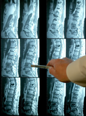
Patients often have the misconception that an MRI scan of their back provides the most comprehensive illustration of the cause of their back pain. They will provide me with a computer disk enclosing their scan and, by no fault of their own, expect that as soon as I view it, I will suddenly know their source of pain. Unfortunately, only in rare cases is an MRI for low back pain really that effective.
The reality is that MRIs are a phenomenal technology that can give valuable information and produce visually stunning images; however, in practice, specifically for low back pain, they are of little value and are notorious for misleading findings.
After an MRI scan, patients will become understandably anxious, more than likely having heard a diagnosis such as, “Disc Herniation” or the words, “bulge” and “protrusion”. While your diagnosis may in fact be correct, remember that NOT ALL LUMBAR DISC HERNIATION’S CAUSE PAIN. In other words, even if your diagnosis is correct, the source of your pain still is not clear from an MRI scan.
Over the last 20 years, notable studies have proven that people with and without back pain can show abnormal findings on an MRI. To put things in perspective consider this – If we were to gather a group of 100 middle-aged people who never experienced back pain before, you would find approximately 35 people with disc bulges, 18 with protrusions, and 8 with herniation’s. Over 60% would show some form of abnormal findings. Remember – These are people with NO PAIN!
So, can we conclude that MRIs are useless? No, of course not. In many situations, they can be a life saving tool. Primarily, as a tool for surgery, MRIs are critical. But in most cases of low back pain, MRI results can actually be counterproductive.
Let me explain why. Most back pain I see in my office, and most back pain in general, is functional – meaning it is the cause of tight/inhibited muscles, restricted joints and over stressed supporting ligaments – none of which can be visualized on an MRI. Contrarily, what I see on a MRI is structural (e.g. a disc bulge), which often is NOT the cause of pain. However, once something like a disc bulge is identified, patients naturally desire more aggressive treatment targeted to the structural problem and prefer treatments such as steroid injections, pain/nerve blocking medication and even surgery. Once we start down this path it is hard for patients and practitioners to get away from it.
It is definitely easier to define the cause of pain by something you can visually see on a X-ray or MRI, than listen to me say that you have weak core and gluteal muscles that are not providing your spine with enough support. But, once we get bogged down with the idea that there is one underlying abnormal structure, we tend to lose sight of the bigger picture and the overall contributors to your pain.
More often than not, functional causes of back pain are what we should be looking for and the good news is these types of problems respond great to conservative care such as chiropractic, physiotherapy, massage and acupuncture.
The moral of the story is this, do not get hung up on an abnormal MRI findings or the idea that you NEED a MRI to identify the problem. Next time you think your disc bulge is the cause of your pain, get a second opinion to look at other possible causes.
If you are unsure of the cause of your low back pain, please do not hesitate to contact me or stop by the clinic for a second opinion..